


Area of Study at UNE
Biomedical Sciences & Community Health
Preview
Competition Year
2014
Microscope Specs
Confocal Microscope
Image Category
Unaltered Image
Competition Award
Third place
Description
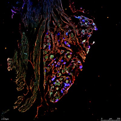
This image characterizes the neuronal profile of a rat dorsal root ganglion (DRG). Retrograde dye (blue) was injected into the proximal periosteum of the tibia and given 7 days to travel retrogradedly along the saphenous nerve to the DRG. The tissue was removed and immunohistochemistry performed; neurons were stained red with mu-opioid receptor antibody and green with IB4 antibody. Quantitative analysis is then performed to determine the amount of neuronal cells that are colocalized (blue with green or red).
Image Creation Date
12-2013